


Perfect Sight Without Glasses
by William H. Bates, M. D. Д. Бейтс
PERFECT SIGHT WITHOUT GLASSES
CHAPTER V
THE TRUTH ABOUT ACCOMMODATION AS DEMONSTRATED BY A STUDY OF IMAGES REFLECTED FROM THE LENS, CORNEA, IRIS AND SCLERA
AS the conclusions to which the experiments described in the preceding chapter pointed were diametrically opposed to those reached by Helmholtz in his study of the images reflected from the front of the lens, I determined to repeat the experiments of the German investigator and find out, if possible, why his results were so different from my own. I devoted four years to this work, and was able to demonstrate that Helmholtz had erred through a defective technique, the image obtained by his method being so variable and uncertain that it lends itself to the support of almost any theory.
I worked for a year or more with the technique of Helmholtz, but was unable to obtain an image from the front of the lens which was sufficiently clear or distinct to be measured or photographed. With a naked candle as the source of light a clear and distinct image could be obtained on the cornea; on the back of the lens it was quite clear; but on the front of the lens it was very imperfect. Not only was it blurred, just as Helmholtz stated, but without any ascertainable cause it varied greatly in size and intensity. At times no reflection could be obtained at all, regardless of the angle of the light to the eye of the subject, or of the eye of the observer to that of the subject. With a diaphragm I got a clearer and more constant image, but it still was not sufficiently reliable to be measured. To Helmholtz the indistinct image of a naked flame seemed to show an appreciable change, while the images obtained by the aid of the diaphragm showed it more clearly; but I was unable, either with a diaphragm or without it, to obtain images which I considered sufficiently distinct to be reliable.
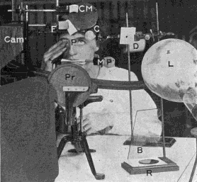
Fig. 24. Arrangements for Photographing Images Reflected From the Eyeball
CM, concave mirror in which the subject may observe the images reflected from various parts of her eye; C, condenser; D, diaphragm; L, 1000-watt lamp; F, forehead rest; MP, bar which the subject grasps with her teeth for the purpose of holding her head steady- P, plane mirror upon which is pasted a letter of diamond type and in which is reflected a Snellen test card twenty feet behind the subject (the mirror is just above theletter P); CAM, camera; Pr, perimeter used to measure the angle of the light to the eye; R, plane mirror reflecting light from the 1000-watt lamp upon the eye, which otherwise would be in total darkness except for the part from which the highly condensed image of the filament is reflected; B, blue glass screen used to modify the light reflected from the mirror R. When the subject read the bottom line of the Snellen test card reflected in themirror P her eye was at rest, and when she saw the letter of diamond type distinctly it was accommodated ten diopters, as demonstrated by the retinoscope.
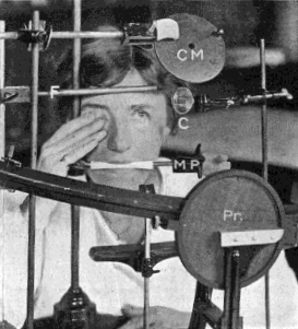
Fig. 25. Arrangements for Holding the Head of the Subject Steady While Images Were Being Photographed
CM, concave mirror; F, forehead rest; C, condenser; MP, mouthpiece; Pr, perimeter.
Men who had been teaching and demonstrating Helmholtz's theory repeated his experiments for my benefit; but the images which they obtained on the front of the lens did not seem to me any better than my own. After studying these images almost daily for more than a year I was unable to make any reliable observation regarding the effect of accommodation upon them. In fact, it seemed that an infinite number of appearances might be obtained on the front of the lens when a candle was used as the source of illumination. At times the image became smaller during accommodation and seemed to sustain the theory of Helmholtz; but just as frequently it became larger. At other times it was impossible to tell what it did.
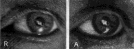
Fig. 26. Image of Electric Filament on the Front of the Lens
R, rest; A, accommodation. Under the magnifying glass no change can be observed in the size of the two images. The image at the right lookslarger only because it is more distinct. To support the theory of Helmholtz it ought to be the smaller. The comet's tail at the left of the two images is an accidental reflection from the cornea. The spot of light beneath is a reflection from the light used to illuminate the eye while the photographswere being taken. It took two years to get these pictures.
With a thirty-watt lamp, a fifty-watt lamp, a 250-watt lamp and a 1000-watt lamp, there was no improvement. The light of the sun reflected from the front of the lens produced an image just as cloudy and uncertain as the reflections from other sources of illumination, and just as variable in shape, intensity and size. To sum it all up, I was convinced that the anterior surface of the lens was a very poor reflector of light, and that no reliable images could be obtained from it by the means described.
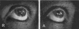
Fig. 27. Images of the Electric Filament Reflected Simultaneously From the Cornea and Lens
R, rest; A, accommodation. The size of the images in both pictures is the same. The corneal image is so small that it has not been noticeablyaltered by the slight change that takes place in the cornea during accommodation. In A both images have changed their position and the endof the reflection from the lens has been cut off by the iris, but its width remains the same. The white spot between the two images of the filamentis a reflection from the lamp used to illuminate the eye. Note that in A more of the sclera is visible, owing to the elongation of the eyeball duringaccommodation.
After a year or more of failure I began to work at an aquarium on the eyes of fish. It was a long story of failure. Finally I became able, with the aid of a strong light—1000 watts—diaphragm with a small opening and a condenser, to obtain, after some difficulty, a clear and distinct image from the cornea of fish. This image was sufficiently distinct to be measured, and after many months a satisfactory photograph was obtained. Then the work was resumed on the eyes of human beings. The strong light, combined with the diaphragm and condenser, the use of which was suggested by their use to improve the illumination of a glass slide under the microscope, proved to be a decided improvement over the method of Helmholtz, and by means of this technique an image was at last obtained on the front of the lens which was sufficiently clear and distinct to be photographed. This was the first time, so far as published records show, that an image of any kind was ever photographed from the front of the lens. Professional photographers whom I consulted with a view to securing their assistance assured me that the thing could not be done, and declined to attempt it. I was therefore obliged to learn photography, of which I had previously known nothing, myself, and I then found that so far as the image obtained by the method of Helmholtz is concerned the professionals were right.
The experiments were continued until, after almost four years of constant labor, I obtained satisfactory pictures before and after accommodation and during the production of myopia and hypermetropia, not only of images on the front of the lens, but of reflections from the iris, cornea, the front of the sclera (white of the eye) and the side of the sclera. I also became able to obtain images on any surface at will without reflections from the other parts. Before these results were obtained, however, many difficulties had still to be overcome.
Complicating reflections were a perpetual source of trouble. Reflections from surrounding objects were easily prevented; but those from the sides of the globe of the electric light were difficult to deal with, and it was useless to try to obtain images on the front of the lens until they had been eliminated, or reduced to a minimum, by a proper adjustment of the light. The same apparent adjustment did not, however, always give similar results. Sometimes there would be no reflections for days; then would come a day when, with the light apparently at the same angle, they would reappear.
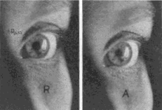
Fig. 28. Image of Electric Filament Upon the Cornea
R, rest; A, accommodation. The image is smaller in A, but the change is so slight as to be scarcely noticeable, showing that the alteration in the shape of the cornea during accommodation is very slight. For this reason the ophthalmometer, with its small image, has been thought to demonstrate that the cornea did not change during accommodation.
With some adjustments of the light multiple images were seen reflected from the front of the lens. Sometimes these images were arranged in a horizontal line, sometimes in a vertical one and sometimes at angles of different degrees, while their distance from each other also varied. Usually there were three of them; sometimes there were more; and sometimes there were only two. Occasionally they were all of the same size, but usually they varied, there being apparently no limit to their possibilities of change in this and other respects. Some of them were photographed, indicating that they were real reflections. Changes in the distance of the diaphragm from the light and from the condenser, and alterations in the size and shape of its opening, appeared to make no difference. Different adjustments of the condenser were equally without effect. Changes in the angle at which the light was adjusted sometimes lessened the number of images and sometimes increased them, until at last an angle was found at which but one image was seen. The images appear, in fact, to have been caused by reflections from the globe of the electric light.
Even after the light had been so adjusted as to eliminate reflections it was often difficult, or impossible, to get a clear and distinct image of the electric filament upon the front of the lens. One could rearrange the condenser and the diaphragm and change the axis of fixation, and still the image would be clouded or obscured and its outline distorted. The cause of the difficulty appeared to be that the light was not adjusted at the best angle for the purpose and it was not always possible to determine the exact axis at which a clear, distinct image would be produced. As in the case of the reflections from the sides of the globe, it seemed to vary without a known cause. This was true, however: that there were angles of the axis of the globe which gave better images than others, and that what these angles were could not be determined with exactness. I have labored with the light for two or three hours without finding the right angle. At other times the axis would remain unchanged for days, giving always a clear, distinct image.
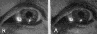
Fig. 29. Image of Electric Filament on the Front of the Sclera
R, rest; A, accommodation. During accommodation the front of the sclera becomes more convex, because the eyeball has elongated, just as a camera is elongated when it is focussed upon a near object. The spot of light on the cornea is an accidental reflection.
The results of these experiments confirmed the conclusions drawn from the previous ones, namely, that accommodation is due to a lengthening of the eyeball, and not to a change in the curvature of the lens. They also confirmed, in a striking manner, my earlier conclusions as to the conditions under which myopia and hypermetropia are produced.1
The images photographed from the front of the lens did not show any change in size or form during accommodation. The image on the back of the lens also remained unchanged, as observed through the telescope of the ophthalmometer; but as there is no dispute about its behavior during accommodation, it was not photographed. Images photographed from the iris before and during accommodation were also the same in size and form, as was to be expected from the character of the lens images. If the lens changed during accommodation, the iris, which rests upon it, would change also.
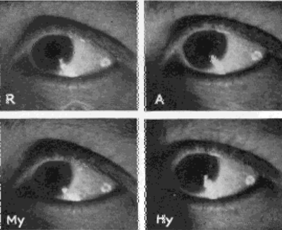
Fig. 30. Images on the Side of the Sclera
R, rest; A, accommodation. The image in A is the larger, indicating a flattening of the side of the sclera as the eyeball elongates. My, Myopia. The eye is straining to see at the distance and the image is larger, indicating that the eyeball has elongated, resulting in a flattening of the side of the sclera. Hy, Hypermetropia. The eye is straining to see at two inches. The image is the smallest of the series, indicating that the eyeball has become shorter than in any of the other pictures, and the side of the sclera more convex. The two lower pictures confirm the author's previous observations that farsight is produced when the eye strains to see near objects and nearsight when it strains to see distant objects.
The images photographed from the cornea and from the front and side of the sclera showed, however, a series of four well-marked changes, according to whether the vision was normal or accompanied by a strain. During accommodation the images from the cornea were smaller than when the eye was at rest, indicating elongation of the eyeball and a consequent increase in the convexity of the cornea. But when an unsuccessful effort was made to see at the near-point, the image became larger, indicating that the cornea had become less convex, a condition which one would expect when the optic axis was shortened, as in hypermetropia. When a strain was made to see at a distance the image was smaller than when the eye was at rest, again indicating elongation of the eyeball and increased convexity of the cornea.
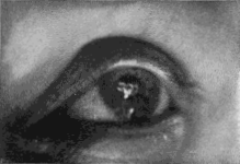
Fig. 31. Multiple Images Upon the Front of the Lens
This picture illustrates one of the difficulties that had to be overcome in photographing images reflected from various parts of the eyeball. Unless the light was adjusted at precisely the right angle the filament was multiplied by reflection from the sides of the globe. Usually the image was doubled, sometimes it was tripled, as shown in the picture, and sometimes it was quadrupled. Often days of labor were required to eliminate these reflections, and for reasons that were not definitely determined the same adjustment did not always give the same results Sometimes all would go well for days, and then, without any apparent reason, the multiple images would return.
The images photographed from the front of the sclera showed the same series of changes as the corneal images, but those obtained from the side of the sclera were found to have changed in exactly the opposite manner, being larger where the former were smaller and vice versa, a difference which one would naturally expect from the fact that when the front of the sclera becomes more convex the sides must become flatter.
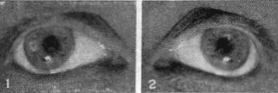
Fig. 32. Reflection of the Electric Filament From the Iris
This picture is shown to illustrate the fact that it is possible to get a reflection from any reflecting surface of the eyeball without reflections from the other parts, although these may be exposed. This is done by changing the angle of the light to the eye. In No. 1 observations of the eye at the time the picture was taken demonstrated that the image was from the iris, not from the cornea, and the fact is also apparent in the picture. (Compare the image with the corneal reflection in Fig. 28.) In No. 2, where the image overlaps the margin of the pupil, the fact that the reflection is from the iris is manifest from the circumstance that only part of the filament is seen. If it were from the cornea, the whole of it would be reflected. Note in this picture that there is no reflection from the lens. The images on the iris did not change their size or shape during accommodation, demonstrating again that the lens, upon which the iris rests, does not change its shape when the eye adjusts itself for near vision.
When an effort was made to see at a distance the image reflected from the side of the sclera was larger than the image obtained when the eye was at rest, indicating that this part of the sclera had become less convex or flatter, because of elongation of the eyeball. The image obtained during normal accommodation was also larger than when the eye was at rest, indicating again a flattening of the side of the sclera. The image obtained, however, when an effort was made to see near was much smaller than any of the other images, indicating that the sclera had become more convex at the side, a condition which one would expect when the eyeball was shortened, as in hypermetropia.
The most pronounced of the changes were noted in the images reflected from the front of the sclera. Those on the side of the sclera were less marked, and, owing to the difficulty of photographing a white image on a white background, could not always be readily seen on the photographs. They were always plainly apparent, however, to the observer, and still more so to the subject, who regarded them in a concave mirror. The alterations in the size of the corneal image were so slight that they did not show at all in the photographs, except when the image was large, a fact which explains why the ophthalmometer, with its small image, has been thought to show that the cornea did not change during accommodation. They were always apparent, however, to the subject and observer.
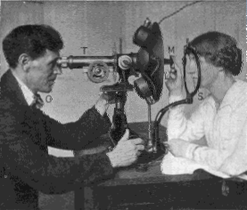
Fig. 33. Demonstrating That the Back of the Lens Does Not Change During Accommodation
The filament of an electric light (L) is shining into the eye of the subject (S), and the reflection on the back of the lens can be seen by the observer (O) in the telescope (T). The subject holds in her hand, at a distance of four inches, a mirror on which is pasted a small letter, and in which is reflected a Snellen test card hung above and behind her head at a distance of twenty feet. The retinoscope reveals that when she looks at the reflection of the test card and reads the bottom line the eye is at rest, and that when she looks at the letter pasted on the mirror it accommodates. The image on the lens does not change during these changes of focus. The telescope is the telescope of the ophthalmometer, the prisms having been removed. As there is no dispute about the behavior of the back of the lens during accommodation this image was not photographed.
The corneal image was one of the easiest of the series to produce and the experiment is one which almost anyone can repeat, the only apparatus required being a fiftycandlepower lamp—an ordinary electric globe—and a concave mirror fastened to a rod which moves back and forth in a groove so that the distance of the mirror from the eye can be altered at will. A plane mirror might also be used; but the concave glass is better, because it magnifies the image. The mirror should be so arranged that it reflects the image of the electric filament on the cornea, and so that the eye of the subject can see this reflection by looking straight ahead. The image in the mirror is used as the point of fixation, and the distance at which the eye focuses is altered by altering the distance of the mirror from the eye. The light can be placed within an inch or two of the eye, as the heat is not great enough to interfere with the experiment. The closer it is the larger the image, and according to whether it is adjusted vertically, horizontally, or at an angle, the clearness of the reflection may vary. A blue glass screen can be used, if desired, to lessen the discomfort of the light. If the left eye is used by the subject—and in all the experiments it was found to be the more convenient for the purpose—the source of light should be placed to the left of that eye and as much as possible to the front of it, at an angle of about forty-five degrees. For absolute accuracy the light and the head of the subject should be held immovable, but for demonstration this is not essential. Simply holding the bulb in his hand the subject can demonstrate that the image changes according to whether the eye is at rest, accommodating normally for near vision, or straining to see at a near or a distant point.
In the original report were described possible sources of error and the means taken to eliminate them.
1. Bates: The Cause of Myopia, N. Y. Med. Jour., March 16, 1912. [link]
| Уход за глазамиФизикаНаука БейтсаЛазер. коррек.Синя. под глаз.Зуд, жжения в глазахВраче. тайнаДр. болезни | |