


Perfect Sight Without Glasses
by William H. Bates, M. D. Д. Бейтс
PERFECT SIGHT WITHOUT GLASSES
CHAPTER II
SIMULTANEOUS RETINOSCOPY
MUCH of my information about the eyes has been obtained by means of simultaneous retinoscopy. The retinoscope is an instrument used to measure the refraction of the eye. It throws a beam of light into the pupil by reflection from a mirror, the light being either outside the instrument—above and behind the subject—or arranged within it by means of an electric battery. On looking through the sight-hole one sees a larger or smaller part of the pupil filled with light, which in normal human eyes is a reddish yellow, because this is the color of the retina, but which is green in a cat's eye, and might be white if the retina were diseased. Unless the eye is exactly focussed at the point from which it is being observed, one sees also a dark shadow at the edge of the pupil, and it is the behavior of this shadow when the mirror is moved in various directions which reveals the refractive condition of the eye. If the instrument is used at a distance of six feet or more, and the shadow moves in a direction opposite to the movement of the mirror, the eye is myopic. If it moves in the same direction as the mirror, the eye is either hypermetropic or normal; but in the case of hypermetropia the movement is more pronounced than in that of normality, and an expert can usually tell the difference between the two states merely by the nature of the movement. In astigmatism the movement is different in different meridians. To determine the degree of the error, or to distinguish accurately between hypermetropia and normality, or between the different kinds of astigmatism, it is usually necessary to place a glass before the eye of the subject. If the mirror is concave instead of plane, the movements described will be reversed; but the plane mirror is the one most commonly used.
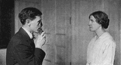
Fig. 8. The Usual Method of Using the Retinoscope
The observer is so near the subject that the latter is made nervous, and this changes the refraction.
This exceedingly useful instrument has possibilities which have not been generally realized by the medical profession. Most ophthalmologists depend upon the Snellen test card, supplemented by trial lenses, to determine whether the vision is normal or not, and to determine the degree of any abnormality that may exist. This is a slow, awkward and unreliable method of testing the vision, and absolutely unavailable for the study of the refraction of the lower animals, of infants, and of adult human beings under the conditions of life.
The test card and trial lenses can be used only under certain favorable conditions, but the retinoscope can be used anywhere. It is a little easier to use it in a; dim light than in a bright one, but it may be used in any light, even with the strong light of the sun shining directly into the eye. It may also be used under many other unfavorable conditions.
It takes a considerable time, varying from minutes to hours, to measure the refraction with the Snellen test card and trial lenses. With the retinoscope, however, it can be determined in a fraction of a second. By the former method it would be impossible, for instance, to get any information about the refraction of a baseball player at the moment he swings for the ball, at the moment he strikes it, and at the moment after he strikes it. But with the retinoscope it is quite easy to determine whether his vision is normal, or whether he is myopic, hypermetropic, or astigmatic, when he does these things; and if any errors of refraction are noted, one can guess their degree pretty accurately by the rapidity of the movement of the shadow.
With the Snellen test card and trial lenses conclusions must be drawn from the patient's statements as to what he sees; but the patient often becomes so worried and confused during the examination that he does not know what he sees, or whether different glasses make his sight better or worse; and, moreover, visual acuity is not reliable evidence of the state of the refraction. One patient with two diopters of myopia may see twice as much as another with the same error of refraction. The evidence of the test card is, in fact, entirely subjective; that of the retinoscope is entirely objective, depending in no way upon the statements of the patient.
In short, while the testing of the refraction by means of the Snellen test card and trial lenses requires considerable time, and can be done only under certain artificial conditions, with results that are not always reliable, the retinoscope can be used under all sorts of normal and abnormal conditions on the eyes both of human beings and the lower animals; and the results, when it is used properly, can always be depended upon. This means that it must not be brought nearer to the eye than six feet; otherwise the subject will be made nervous, the refraction, for reasons which will be explained later, will be changed, and no reliable observations will be possible. In the case of animals it is often necessary to use it at a much greater distance.
For thirty years I have been using the retinoscope to study the refraction of the eye. With it I have examined the eyes of tens of thousands of school children, hundreds of infants and thousands of animals, including cats, dogs, rabbits, horses, cows, birds, turtles, reptiles and fish. I have used it when the subjects were at rest and when they were in motion—also when I myself was in motion; when they were asleep and when they were awake or even under ether and chloroform. I have used it in the daytime and at night, when the subjects were comfortable and when they were excited; when they were trying to see and when they were not; when they were lying and when they were telling the truth; when the eyelids were partly closed, shutting off part of the area of the pupil, when the pupil was dilated, and also when it was contracted to a pin-point; when the eye was oscillating from side to side, from above downward and in other directions. In this way I discovered many facts which had not previously been known, and which I was quite unable to reconcile with the orthodox teachings on the subject. This led me to undertake the series of experiments already alluded to. The results were in entire harmony with my previous observations, and left me no choice but to reject the entire body of orthodox teaching about accommodation and errors of refraction. But before describing these experiments I must crave the readers patience while I present a resume of the evidence upon which the accepted views of accommodation are based. This evidence, it seems to me, is as strong an argument as any I could offer against the doctrine that the lens is the agent of accommodation, while an understanding of the subject is necessary to an understanding of my experiments.
1. Herman Snellen (1835-1908). Celebrated Dutch ophthalmologist, professor of ophthalmology in the University of Utrecht and director of the Netherlandic Eye Hospital. The present standards of visual acuity were proposed by him, and his test types became the model for those now in use.
| Уход за глазамиФизикаНаука БейтсаЛазер. коррек.Синя. под глаз.Зуд, жжения в глазахВраче. тайнаДр. болезни | |